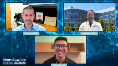
From Imaging to Biomarkers: Improving Remyelination Assessment in MS
Neurologists Ari Green, MD, and Paul Tesar, PhD, discuss the current limitations of remyelination assessment tools in MS and highlight efforts to develop more reliable clinical measures.
Episodes in this series

Despite major advancements in the treatment of multiple sclerosis (MS), including the emergence of highly effective disease-modifying therapies that target inflammation and reduce relapse rates, there remains a critical unmet need: the ability to repair damaged myelin. Remyelination—the process by which the central nervous system regenerates myelin sheaths around axons—is a key neuroprotective and potentially restorative goal in MS. While spontaneous remyelination can occur, particularly early in the disease, it is often incomplete and declines over time, contributing to irreversible neurological disability.
In recent years, an influx of research has significantly advanced our understanding of the biology of remyelination, uncovering new molecular targets, cellular mechanisms, and imaging modalities. Yet, despite promising results in preclinical models, there are still no FDA-approved therapies that directly promote remyelination. In this panel discussion, Ari Green, MD, and Paul Tesar, PhD, examine the evolving landscape of remyelination science—highlighting the translational challenges, trial design hurdles, and promising agents in development—while offering insights into the future of regenerative strategies in MS care.
In this episode, Green, co-director of the Innovation Program for Repair and Remyelination at UCSF, and Tesar, director of the Institute for Glial Sciences at Case Western University School of Medicine, explore how remyelination is currently assessed in multiple sclerosis clinical trials and why measuring myelin repair remains a challenge. They discuss the limitations of imaging and functional tools, the need for more direct markers of myelin restoration, and the risk of overinterpreting early data. The conversation also highlights the emerging role of biomarkers and why validating assessment methods depends heavily on the success of future remyelinating therapies.
Transcript was edited for clarity.
Marco Meglio: Let’s move on to how we’re currently assessing remyelination in clinical trials, and what’s being done to improve those methods.
Ari Green, MD: This is a critical question for the field. Across medicine, the only way to develop tools that reliably assess whether something works is to actually test drugs that modulate disease—and then measure whether your hypothesis holds true. It's an iterative process. Through clinical trials, we've identified several tools that may be useful. These include functional measures of electrical signal transmission in the brain—since myelin enables faster conduction—as well as long-term markers of axon survival. Imaging is another area of interest, though we have to be careful in interpreting what these techniques really measure.
I’ll give you an example: A recent paper suggested that people who run marathons experience brain demyelination during the run and then remyelinate afterward. Now, I want to give the authors credit, but for those of us in the myelin field, that conclusion is implausible. It illustrates the risk of misinterpreting imaging tools that might reflect myelin, but also reflect other biological changes. We have to validate these methods rigorously, aligning them with what we know about myelin biology from detailed research.
Paul Tesar, PhD: Ari’s right—MRI-based imaging is valuable but remains an indirect measure of remyelination. There's room to improve its specificity and sensitivity, whether through high-resolution MRI or PET tracers that might more directly visualize myelin. Functional assessments like visual evoked potentials are currently the field standard, but they only capture remyelination in the visual system. We need broader tools.
Another emerging area is biomarkers—could there be blood or CSF proteins that reflect oligodendrocyte regeneration? For instance, unlike markers like neurofilament light chain, which indicate axonal injury, we might be able to detect something specific to new oligodendrocyte formation. Since these cells are long-lived, any new ones would stand out against a low background signal. Even circulating RNAs could hold promise. But as Ari said, until we have an effective remyelinating therapy, it's hard to know which assessment tools truly correlate with clinical benefit.
Newsletter
Keep your finger on the pulse of neurology—subscribe to NeurologyLive for expert interviews, new data, and breakthrough treatment updates.


