Feature
Article
The Evolutionary Arch of Stereoelectroencephalography in the United States
Author(s):
Key Takeaways
- SEEG provides a less invasive approach for identifying epileptogenic networks, crucial for complex epilepsy cases and failed prior surgeries.
- Advancements in imaging, robotic systems, and electrode technologies have enhanced SEEG's effectiveness in surgical decision-making.
SEEG has transformed the surgical investigation of refractory epilepsy, enabling clinicians to map complex seizure networks with precision, leading to more targeted treatment options for patients.
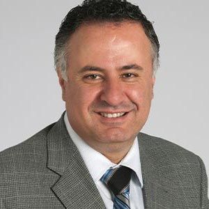
Demitre Serletis, MD, PhD

Stereoelectroencephalography (SEEG) is an important surgical method used in the investigation of medically refractory epilepsy, to identify the networks involved in early seizure organization and propagation. SEEG was originally conceptualized by the French school of epileptology in the 1950s, and made its first appearance in the United States nearly 50 years later, following its inception at Cleveland Clinic on March 24, 2009. To date, our institution has now performed more than 1000 SEEG procedures for patients with intractable epilepsy, using the method to provide critical insights into some of the most challenging epilepsy cases, including those patients who had previously failed surgical intervention(s). The method has become the crux of epilepsy surgical decision-making, offering new insights into surgical indications and approaches, procedural advancements, and treatment outcomes.
We focus this review on the evolutionary arch of SEEG since its implementation in the United States, highlighting important lessons learned in regard to patient selection, surgical pre-implantation planning, quantitative SEEG data analysis, and novel SEEG-guided surgical therapies that are emerging on the horizon.
Juan Bulacio, MD

The SEEG Experience at Cleveland Clinic: Past and Present Developments
Initially, when SEEG was first introduced to the United States, the method was primarily indicated for patients with complex epilepsy syndromes, particularly those with suspected focal epilepsy, or failed epilepsy surgery, and in whom noninvasive (and at times invasive) evaluations remained inconclusive. In comparison to the more traditional subdural grid approach for cortical mapping (as was popularized in North America at the time), SEEG offered a comparably less invasive way of recording from deeper cortical and subcortical networks, delivering a spatiotemporal method for recording data directly from the insula, mesial temporal structures, cingulate and posterior orbitofrontal region, amongst other areas. For patients presenting with multifocal or MRI-negative epilepsy, a deeper (and at times more extensive sampling) of potential epileptogenic networks was required, facilitated, and enabled by SEEG.
Surgical techniques continued to evolve, integrating improved image-based guidance, sophisticated robotic stereotactic systems, and a host of new SEEG electrode technologies, each with its own unique advantages and disadvantages. Increasingly more patients who had previously failed prior attempts at surgical resection, or who were initially explored with subdural grid methodology that had failed to localize an epileptogenic zone, suddenly had another surgical option, which led to improved seizure outcomes. Over the past 15 years, the SEEG effect has rippled out across the United States (and the rest of the world), with additional centers taking renewed interest in adopting the method. Important conferences and workshops were organized, including those highly popularized and frequented at Cleveland Clinic (even to this day), to instruct neurologists, neurosurgeons, neuroradiologists, EEG technicians, nuclear imaging specialists, neuropsychologists, and scores of other professionals in the skillset requisite to interpreting, analyzing and performing SEEG.
William Bingaman, MD

As our understanding and expertise in SEEG methodology grow, its applications have expanded, becoming relevant to a wider range of patients, including those with dual pathology, large malformations, or complex brain lesions. A good proportion of patients with intractable epilepsy are now referred for the procedure, which is now regularly used to guide tailored surgical resections and laser ablations. SEEG also offers the ability to perform localized radiofrequency ablations along specific electrode trajectories, helping to serve as a crucial diagnostic interrogation of tentative epileptogenic networks that may be in question, prior to any surgical intervention being offered. The method continues to play an increasingly important role in the decision to offer neuromodulation, whether by responsive neurostimulation (RNS) or deep brain stimulation (DBS). These surgical options continue to benefit from an informed, data-driven approach conferred by SEEG methodology. Finally, when SEEG fails to localize the epileptogenic network, it offers an important ‘hard stop’ for further surgical attempts that are unlikely to help the patient.
Lessons Learned from Patient Selection and Preimplantation Planning
Through our experience with more than 1000 patients undergoing SEEG as part of their epilepsy work-up, several key lessons have emerged regarding patient selection and preimplantation planning. The decision to offer SEEG is made at a multidisciplinary Patient Management Conference (PMC), whereby all data and clinical information are reviewed. It is at these sessions that critical decisions are made regarding patient selection and candidacy for SEEG, and once the decision is made to proceed, to determine the number and intended trajectory of each electrode. These conferences are imperative to safe surgical decision-making and effective planning and always include an ethicist to ensure the patient’s best interests are well represented by the team. Increased numbers of patients with different types of epilepsy have been reviewed for their candidacy for SEEG, including patients harboring challenging brain lesions such as temporal encephaloceles; polymicrogyrias; periventricular nodular heterotopias; and large hemispheric malformations or lesions that were previously deemed inoperable. SEEG has been essential to surgical decisions around these pathologies, including offering hope to those patients with MR-negative findings, with good-to-excellent results now published in the literature.
Imad Najm, MD

Meticulous analysis of patient outcomes has also highlighted the importance of individualized pre-implantation planning, reliant on multimodal review of MRI, PET, SPECT (single-photon emission computed tomography), and MEG (magnetoencephalography) imaging, amongst other modalities. These noninvasive methods help to define the extent of a given lesion—when visible—and its relationship to functional networks of the brain. Novel post-processing advances in MRI, including morphometric analysis and MR-fingerprinting, offer supplementary new ways to identify changes in brain tissue using multiparametric quantitative imaging, that may be used to precisely identify a tentative epileptogenic region. These results are collectively and comprehensively reviewed by the team at PMC, generating the localization hypotheses to be tested and the networks to be assessed. The PMC discussions guide the careful process of preimplantation planning and allow for multidisciplinary group-informed selection of SEEG trajectories (including targets and entry sites) of importance.
Another significant lesson herein stems from our experience with patients who previously did not receive SEEG. In these cases, it was observed that the utilization of SEEG could have further refined and tailored the surgical approach in a large number of resective candidates, particularly by identification of the precise location of epileptogenic foci while concomitantly minimizing the risk of post-operative functional deficits.
Data Analysis, Surgical Decision-Making, and Future Technologies
Analysis of SEEG data has proven to be instrumental in guiding surgical decisions regarding resection, ablation, and/or neuromodulation. In our practice, a clear framework has been established to analyze interictal and ictal data, correlating these findings with clinical seizure semiology and the patient’s anatomy, thereby comprising an “anatomo-electro-clinical” analysis of the epileptogenic network(s) in question. Traditionally, the review of SEEG data is performed by clinician experts. Nevertheless, with increased sophistication of computational methods, bolstered by advances in mathematics, physics, neuroengineering, signal processing, and data-driven science (including artificial intelligence and machine learning), there are increasingly new methods emerging for the investigation of nonlinear dynamical complexity and related signal biomarkers that are inherent to in vivo spatiotemporal SEEG recordings and brain state transitions. These quantitative approaches offer exciting new insights into the identification and localization of epileptogenic networks, leading to even more targeted, precision-based surgical decision-making for epilepsy patients.
Hence, the future of SEEG-guided epilepsy treatment holds great promise, particularly with continued advancements in imaging technologies and data analysis techniques. Enhanced pre-implantation planning through improved modalities, including magnetic resonance fingerprinting (MRF), will facilitate an improved identification of subtle lesions, their underlying pathology, and intrinsic epileptogenicity, leading to more precise SEEG electrode targeting for sampling these regions. Less invasive treatment options will continue to become more prevalent. In particular, the advent and refinement of laser interstitial thermal therapy, high-intensity focused ultrasound, and focused gene therapy represent exciting and potential surgical alternatives, directly informed by the personalized SEEG results for a given patient. These methods have the potential to target epileptogenic networks with minimal disruption to surrounding brain tissue. As mentioned, the integration of artificial intelligence in the post-processing of SEEG data is anticipated to further enhance the ability to identify epileptogenic networks, and thus improve the surgical decision-making and targeting of these regions. Moreover, emerging new quantitative methods to study, interrogate, and even potentially regulate, the spatiotemporal dynamics underlying the transition to seizure events across different brain networks will lead to significant advances in ‘smart’ neuromodulation devices, evolving towards a paradigm featuring personalized stimulation strategies based on real-time seizure detection. The continued development of these approaches paves the way toward a dynamic, exciting, and arguably, more effective approach toward managing intractable epilepsy.
Conclusion
The inception and integration of SEEG into the surgical epilepsy workflow has significantly enriched our understanding and treatment of epilepsy in the United States. The lessons learned from patient selection, pre-implantation planning, and surgical decision-making will undoubtedly shape the future landscape of SEEG-guided epilepsy treatments. With ongoing advancements in imaging, minimally invasive therapies, and intelligent, quantitative data analysis, we are on the cusp of a new era in the management of intractable epilepsy, one that promises improved outcomes for patients across the spectrum of this challenging disorder.
Newsletter
Keep your finger on the pulse of neurology—subscribe to NeurologyLive for expert interviews, new data, and breakthrough treatment updates.


