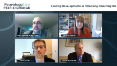
Exciting Developments in Relapsing-Remitting MS
Exciting strategies and biomarkers being investigated to improve the evaluation and treatment of patients with relapsing-remitting multiple sclerosis.
Episodes in this series

Scott D. Newsome, DO, MSCS, FAAN: Looking ahead in the world of MS [multiple sclerosis], as has been alluded to, there are different biomarkers being studied. Maybe we can add these new biomarkers into that category of NEDA [no evidence of disease activity], MIDA [minimal evidence of disease activity], or whatever acronym you want to use for no evidence of disease activity or minimal activity. Bob, tell us about some of the exciting biomarkers that may be used in clinics over the next 5 or so years.
Robert Fox, MD: Ideally, what we want is to be able to look at a patient, look at an MRI, look at a blood test to say,” Based on this, this is the therapy that will work best for you.” At this point, we do not have that. We have markers that predict what [medications] will have problematic safety issues. If the patient has a positive JC [John Cunningham] virus serology, we say stay away from natalizumab. If a patient has a heart arrhythmia, we stay away from the S1P [sphingosine-1-phosphate] modulators. We can use the safety aspects to guide us as to what therapies to stay away from, but we do not know how to be guided toward the more effective therapy for an individual patient.
Once we have chosen a drug and the patient starts it, MRI is the key biomarker right now; it goes along with our clinical assessment of whether the patient is having relapses. What goes along with that is the MRI, and it is usually just the brain MRI because the spinal cord is such an eloquent pathway that, when patients develop lesions in the spinal cord, they can usually tell us about it because they have new symptoms. We get spinal cord imaging now and then for specific reasons, but as a surveillance tool, it is usually relegated to just the brain MRI to identify new lesions.
In terms of blood measures, there are a couple of measures that are looking potentially promising: serum neurofilament light chain [NfL] is probably our most well-developed marker at this point. There are some other markers like serum GFAP [glial fibrillary acidic protein], contactin, and other things that also look promising. Serum neurofilament light chain is the most developed, but at this point, we do not know if that is any better than the MRI. There are some studies that suggest that the serum neurofilament is not as sensitive as the brain MRI in identifying new areas of ongoing inflammation. Although serum neurofilament light chain is widely available, I do not think we yet know how to use it to monitor the disease; what are the cutoffs to say, “That is too much. We need to move on to another therapy.” There is a lot of work being done, and hopefully over the next several years, we will have a better way of being able to detect ongoing inflammation despite our current therapies.
Scott D. Newsome, DO, MSCS, FAAN: I am glad you brought up some of the unknowns about NfL because some of the things I am worried about due to the lack of data are these questions: what is the impact of comorbidities and what is the impact of age on NfL? We do not have a lot of data, especially within the MS population. There is more to come.
The one message that I will usually say to trainees is that, if we have serum neurofilament light chain available, do not just take it at 1 time point. This is something that you are going to have to take at multiple points just like we do with the MRI, and just like we do in the clinic when we see people over time. It is an evolving story.
Clyde, what do you think? Are there any other cool things out there? I know at the NIH [National Institutes of Health], they are doing a lot of interesting imaging work. You mentioned central vein sign a couple of times. What do you think?
Clyde E. Markowitz, MD: To add to the neurofilament light chain conversation, we might be able to use it as a cross-sectional idea where, if you are evaluating somebody from the beginning and get a neurofilament light chain level, then that may give you some information that is going to tell you this is a high-risk individual you are going to have to worry about going forward. We also know that, on many of the clinical trials, the investigators have been using this biomarker for the idea of response to therapy. If somebody has a good response, where you follow them longitudinally and their levels came down, you might feel more comfortable that this is a good responder. In reality, we will probably incorporate it into our world. I do not think it will be able to replace things, but it could be an additional piece of data that will help us with all the other metrics.
OCT [optical coherence tomography] is another thing that is interesting, where we have been looking at that for probably 15, 20 years by now. We think that the back of the eye, being purely neuronal, could be a marker of what is happening in the central nervous system, particularly within the brain. There are now some data to suggest that it correlates fairly well with MRI. It may even correlate to some degree with EDSS [Expanded Disability Status Scale] scores. We are now starting to see some data that suggest good correlation with progression of the disease. It could be worth considering following these people longitudinally with the OCT biomarker.
Looking at MRIs, the newer techniques being implemented, such as central vein sign, become interesting ideas for us to use to figure out whether the lesion is an MS lesion, because as our population ages, there are going to be lesions that show up from hypertension, aging, diabetes, and migraines. Not every white spot on an MRI scan is going to be because of a lack of therapy or an ineffective therapy for MS, because they still have all the other problems that might show up with white matter lesions. Being able to distinguish a white matter lesion that is MS vs a white matter lesion from hypertension can be helpful in terms of trying to figure out a decent response to whatever therapy you are going to consider.
There is a lot going on with high field magnets and being able to look at cortical lesions. The whole arena of these assessments is just starting to scratch the surface now where we may be able to say that we have abnormalities on imaging studies related to thalamic volumes or gray matter volumes: things that are going to give us good windows into the parts of MS that we do not adequately assess currently. How is somebody functioning in their day-to-day world in their job? Do they have complaints of fatigue or do they have the brain fog that they all complain about? We are not capturing that in most of our clinical trials, but some of the research studies are pointing us in the direction of how we can get at that and be able to follow that over time. Higher field strength magnets looking at spinal cord imaging is going to be better than what we currently have. The field going forward is starting to get into this biomarker arena that is evolving, and we will be able to care for our patients even better over the next 5 to 10 years.
Scott D. Newsome, DO, MSCS, FAAN: Thank you for watching this NeurologyLive® Peer Exchange. If you enjoyed the content, which I hope you did, please subscribe to our e-newsletters to receive up-and-coming Peer Exchanges and other great content right in your inbox.
Transcript Edited for Clarity
Newsletter
Keep your finger on the pulse of neurology—subscribe to NeurologyLive for expert interviews, new data, and breakthrough treatment updates.

