
At SLEEP 2026, the head of Orexin Franchise Development and Neuroscience Programs at Takeda discussed new phase 3 data of oveporexton in patients with narcolepsy type 1. [WATCH TIME: 5 minutes]
At SLEEP 2026, the head of Orexin Franchise Development and Neuroscience Programs at Takeda discussed new phase 3 data of oveporexton in patients with narcolepsy type 1. [WATCH TIME: 5 minutes]

At SLEEP 2026, the research intern at Beth Israel Deaconess Medical Center described the development of an electronic health record–based machine learning model for obstructive sleep apnea screening. [WATCH TIME: 3 minutes]
Understanding how amyloid pathology develops years before symptoms emerge is essential to appreciating the growing role of blood-based biomarkers in earlier Alzheimer disease detection.
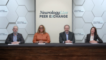
In this episode titled "MS and Pregnancy: Addressing Misperceptions About Anti-CD20 Therapies and Childbearing," the panel addresses common patient misperceptions about anti-CD20 therapies. Dr. Williams begins with a topic Dr. Bove is particularly well-positioned to address: the misperception that B-cell depleting therapies are unsafe or incompatible with pregnancy.
As anti-CD20 therapies become entrenched in the treatment paradigm for multiple sclerosis (MS), questions about their long-term use, sequencing, and broader impact on patient care are moving to the forefront.
In episode titled "Choosing Among Anti-CD20 Therapies in MS: Patient Profiles and Clinical Decision Making," Dr. Williams asks Dr. Stephen Krieger whether specific patient profiles favor one anti-CD20 agent over another among ocrelizumab, ofatumumab, and ublituximab.

Neurology News Network for the week ending July 18th, 2026. [WATCH TIME: 4 minutes]
At SLEEP 2026, the sleep medicine specialist at University of Arizona discussed the new American Academy of Sleep Medicine guideline on the use of combination therapy in adults with chronic insomnia. [WATCH TIME: 5 minutes]
At SLEEP 2026, a sleep medicine specialist at Mayo Clinic talked about the 2026 Restless Legs Syndrome Foundation algorithm, recently published in Mayo Clinic Proceedings. [WATCH TIME: 10 minutes]
Mitzi Joi Williams, MD, is joined by Jacquelyn McEwen, MD, and Jacqueline Rosenthal, MD, to discuss the latest updates to MS diagnostic criteria and their impact on diagnosing MS and NMOSD.

The assistant professor in the Department of Behavioral Science at the University of Kentucky discusses a nonpharmacologic intervention designed to address sensory processing impairments in older adults with dementia. [WATCH TIME: 5 minutes]
At AAIC 2026, professor of neurology at Washington University in St. Louis discussed how integrating clinical assessment with amyloid biomarkers can improve diagnostic accuracy in Alzheimer disease. [WATCH TIME: 5 minutes]

At AAIC 2026, associate professor of neurology at Albert Einstein College of Medicine discussed a culinary medicine intervention to improve MIND diet adherence and mood in older adults with type 2 diabetes. [WATCH TIME: 5 minutes]
Neurologist Michio Hirano, MD, provides an overview of thymidine kinase 2 deficiency, discussing its underlying biology, clinical presentation, and natural history before the advent of disease-modifying therapy.

In the largest study of its kind, researchers examined the potential neurological effects of repetitive head impacts in retired elite soccer players, with findings offering new insights into symptoms, brain structure, and future research directions. [WATCH TIME: 3 Minutes]
In this episode titled "Long-Term Management of MS: Monitoring Progression and Supporting Patient Wellness While on Anti-CD20 DMTs," Dr. Williams asks the panel what the clinical conversation looks like for patients who have been on therapy for five to seven or more years — and what new priorities have emerged.
Shared decision-making has become a foundational element of contemporary multiple sclerosis (MS) care, particularly as the number of available disease-modifying therapies has expanded.
This episode discusses "The MS 'Wearing-Off' Phenomenon: Separating Symptom Burden from Immunological Change." Dr. Williams introduces the wearing-off phenomenon — a patient experience in which symptoms feel worse as the next anti-CD20 dose approaches. The panel explores whether this represents a true immunological change or a symptom-level phenomenon.
Ensuring safety over the course of high-efficacy therapy remains a central concern in the management of relapsing multiple sclerosis (MS).

The director of the Division of Neuroscience at IRCCS Istituto Ortopedico Rizzoli discusses why targeting shared biologic pathways, rather than individual mutations, may represent a promising therapeutic strategy for Charcot-Marie-Tooth disease. [WATCH TIME: 2 minutes]

Neurology News Network for the week ending July 11th, 2026. [WATCH TIME: 4 minutes]
At CMSC 2026, the associate professor of neurology at the Medical College of Wisconsin discussed the need to reconsider clinical trial paradigms by recognizing the role of immune cells in MS. [WATCH TIME: 6 minutes]
At CMSC 2026, the director of the multiple sclerosis research unit at Ottawa Hospital discussed how integrating clinical research into MS practice enhances clinician expertise. [WATCH TIME: 5 minutes]
Ugur T. Sener, MD, discusses how findings from the INDIGO trial are influencing treatment timing after surgical resection, including evolving considerations surrounding early intervention, surveillance, fertility, and long-term management.

Matthew Evans, BM, BCh, DPhil, a consultant neurologist at the University of Oxford, discusses the diagnostic approach to small fiber neuropathy and the challenges of confirming immune-mediated disease. [WATCH TIME: 3 minutes]
In this episode titled "Extended Interval Dosing of Anti-CD20 Therapies: Benefits, Risks, and Real-World Experience," moderator Dr. Mitzi Joi Williams introduces the concept of extended interval dosing (EID): rather than dosing on a fixed six-month or monthly schedule, could practitioners dose less frequently while maintaining efficacy — whether for cost, safety, or patient preference?
Williams describes long-term treatment success with ofatumumab in relapsing MS, noting that ASCLEPIOS trial and extension data support sustained reductions in relapse rates, MRI lesions, and confirmed disability progression.
This episode discusses "Hypogammaglobulinemia in MS Patients on Anti-CD20 Therapy: When to Act." Dr. Williams identifies hypogammaglobulinemia — low IgG levels resulting from prolonged B-cell depletion — as one of the most clinically consequential long-term complications of anti-CD20 therapy, and asks the panel how they identify high-risk patients and manage this finding.
Williams explains that at-home, once-monthly ofatumumab is generally well received by patients with relapsing MS, offering convenience for those with active work and family lives while addressing concerns about injections and treatment scheduling.

Neurology News Network for the week ending July 4th, 2026. [WATCH TIME: 4 minutes]